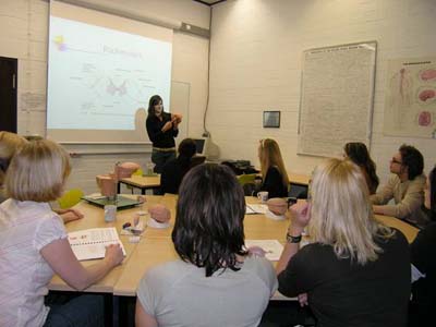
The goal of this course is to educate biology students in basic knowledge of human neuroanatomy including the principal functions of the brain areas.
We wish for students participate actively and literally “grasp” the structures in order to experience the learning of neuroscientific contents in a positive and consolidatory manner. The course consists of two parts which alternate from session to session: 1) “Theory”: Each brain area (7 brain areas in total) is introduced by one or two students.
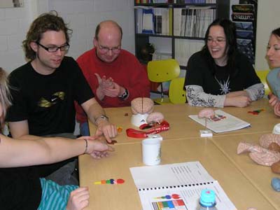
The students learn the general construction, set-up and position of this structure, in addition to their primary function. Students shall model this brain structure with plasticine with the substructures coded in different colors.
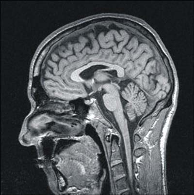
Additionally, students will be given 3D-models of human brains, to study the position of the actual brain structure and its relation to the rest of the brain.
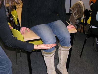
In order to learn the function of the respective brain area, the function will be first demonstrated in small experiments (e.g. patella-reflex for the brain structure “spinal cord”).
Additionally, the neuronal circuits which serve this function are embedded in the plasticine model with plastic pearls (soma) and strings (axons and dendrites).
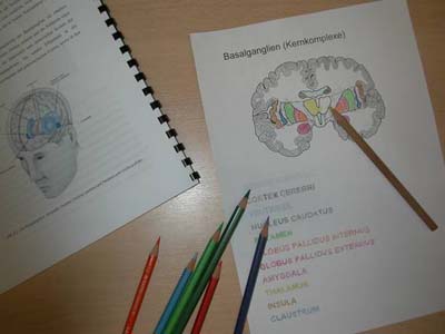
To support self-learning, students will be given black and white line drawings of the brain structure in which they have to identify the substructures and color code them by painting.
Furthermore, the students are able to borrow the 3D brain models to study at home for consolidation.
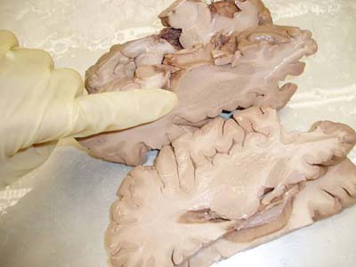
2) “Practice”: The second part of each “brain area” takes place in the Institute of Neuroanatomy.
First, the contents of the previous session will be repeated (set-up and function of the respective brain area) in close interaction with the whole group. Afterwards the students will be given the opportunity to study real conserved brains as well as histological brain slices, to transfer their previously acquired “theoretical” knowledge about the respective brain area to real brain and slices of different scaling,
thereby completing their concept of the human brain.
Additionally, students will learn aspects of malfunctioning brains: effects of lesions, stroke and some mental diseases.
Literature: - Pinel: Biopsychologie, Spektrum Verlag
- Kandel, Schwartz, Jessell: Neurowissenschaften, Spektrum Verlag
- Kandel, Schwartz, Jessell: Principles of Neural Science, McGraw Hill
- Dudel, Menzel, Schmidt: Neurowissenschaften, Springer Verlag
- Kolb, Wilshaw: Neuropsychologie, Spektrum Verlag
- Trepel: Neuroanatomie - Struktur und Funktion, Elsevier Verlag
- Nieuwenhuys, Voogd, van Huijzen: The Human Central Nervous System: A Synopsis and Atlas, Springer Verlag
- Augustine: Human Neuroanatomy, Academic Press
Each student has to prepare one brain structure. At the end of the course an exam is written to test the increase of knowledge. | |